
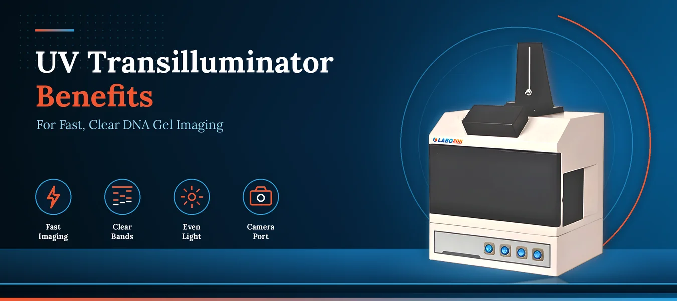
After a gel run ends, the next decision depends on how well the bands can be seen. This guide explains how a UV transilluminator makes DNA fragments visible, where it fits in a laboratory workflow, and which details deserve attention when reviewing instruments such as the UV Trans-illuminator LZ-UVT-A100.
Why Visibility Matters After the Run
Once electrophoresis finishes, a gel looks almost blank. The separated DNA fragments are there, but nothing on the surface tells a technician whether the run worked. A UV transilluminator closes that gap by lighting the gel from below, which makes stained fragments glow so that each band can be examined within moments.
In transilluminator gel electrophoresis work, that quick look decides what happens next. Sharp, evenly lit bands let a team confirm fragment sizes, judge sample quality, and move forward without repeating a run. Faint or uneven bands send laboratories, research centres, and advanced labs back to the bench, which is why the lighting stage deserves as much thought as the gel itself.
From Ultraviolet Light to Visible Bands
The UV transilluminator working principle rests on fluorescence. Ultraviolet lamps sit beneath a filter surface, and the gel rests on top of it. The DNA fragments carry a fluorescent dye, and that dye absorbs the ultraviolet energy and releases it again as visible light. The glow marks exactly where each fragment sits in its lane, so the pattern can be read against a ladder.
Wavelength matters here. The UV Trans-illuminator LZ-UVT-A100 offers a mid-range transmission wavelength of three hundred two nanometers for gel viewing, along with short-range and long-range reflectance wavelengths of two hundred fifty four and three hundred sixty five nanometers. An observation window with an anti-ultraviolet filter lets the operator watch the gel in real time without direct exposure to the light.
Where the Instrument Fits in the Workflow
The UV transilluminator function is simple to state and easy to underestimate: it converts a finished gel into an image that can be interpreted, shared, and stored. In a UV transilluminator gel electrophoresis routine, the gel moves from the tank to the filter surface, the operator checks the banding pattern through the protective window, and a camera records the result for notebooks and reports.
Because the same image is used to decide on next steps and to document the run, even lighting across the full viewing area is more useful than a single bright spot. Lanes at the edge of the gel should read as clearly as lanes in the middle.
Laboratory Uses and Applications
UV transilluminator uses in laboratory settings center on the examination, observation, and photography of electrophoresis gels. The UV transilluminator application appears across molecular biology, genetic engineering, biotechnology, and tissue culture laboratories, where gel images support routine checks and recorded results.
The same UV transilluminator machine can also serve when objects exposed to ultraviolet light need to be photographed through a normal camera, which widens its usefulness beyond gels. Other UV transilluminator uses follow the same pattern: any task that depends on seeing fluorescence under ultraviolet light. Teams weighing these uses against their own workflow can look through the range of transilluminator models to see how light modes and viewing areas differ.
Reading the LZ-UVT-A100 Closely
The UV Trans-illuminator LZ-UVT-A100 pairs uniform lighting with a camera-ready layout. The details below come from its product page, where the full specification table is listed.
Illustration placeholder: instrument with fixed camera port rack.
Even illumination
Ten lamps of six watts each spread light uniformly across the viewing area.
Generous filter area
The transmission filter measures two hundred by one hundred fifty millimeters, leaving room for a full gel.
Fixed camera port
The camera port rack suits almost all SLR cameras, so a normal camera can photograph the glowing gel.
Protected viewing
The anti-ultraviolet observation window filter supports safe, real-time observation.
Durable body
An iron-rust proof painted metal body and long-lasting lamps suit extended use at the bench.
Two light modes
Transmission and reflectance wavelengths cover gel viewing and photography of ultraviolet-exposed objects.
Handling Tips for Clearer Images
Good hardware still depends on good habits. These routines keep images sharp and bands easy to read.
Seat the gel flat
Center the gel on the filter so light reaches it evenly and edge lanes read like middle lanes.
Keep the filter clean
Wipe dust and gel residue from the surface before loading, since debris appears as shadows in a photograph.
Use the protective window
Observe through the anti-ultraviolet window and keep it in place whenever the lamps are on.
Limit lamp time
Switch the lamps on when imaging begins, because extended exposure can fade the dye signal.
Steady the camera
Mount the camera on the fixed port, then adjust exposure rather than moving the camera between shots.
Record the settings
Note camera and exposure settings so a later image can be matched to an earlier one.
Selection Mistakes Worth Avoiding
Judging by lamp output alone
Lamp power says little unless the light is spread evenly across the viewing area.
Overlooking filter size
A gel tray larger than the filter forces repeated repositioning and uneven images.
Ignoring camera access
Without a fixed camera port, image quality depends on handheld positioning.
Skipping viewing protection
Direct ultraviolet exposure during observation is a safety gap, so look for an anti-ultraviolet observation window.
Matching wavelength poorly
A mid-range transmission wavelength suits gel viewing, while short-range and long-range reflectance wavelengths serve other imaging tasks.
Forgetting upkeep and supply
Ask about body finish and lamp service life, and confirm that the voltage and frequency range matches the laboratory supply.
Light Modes and Related Models at a Glance
The two light modes on the LZ-UVT-A100 are built for different jobs. This table shows where they differ.
| Aspect | Transmission mode | Reflectance mode |
|---|---|---|
| Light path | Passes up through the gel from below | Falls on the surface of the object |
| Wavelength | Mid-range, three hundred two nanometers | Short range at two hundred fifty four nanometers, long range at three hundred sixty five nanometers |
| Filter size | Two hundred by one hundred fifty millimeters | Two hundred by eighty millimeters |
| Typical task | Examining and photographing electrophoresis gels | Photographing objects exposed to ultraviolet light |
Laboratories that want a different light source or format can also review the UV Trans-illuminator LZ-UVT-A110, along with the blue light models Blue Light Transilluminator LZ-BLT-A100 and Blue Light Transilluminator LZ-BLT-A101.
The Wider Transilluminator Category
The LZ-UVT-A100 belongs to the transilluminator category, a group of instruments that light a gel from beneath so that separated nucleic acids can be seen and photographed. The group includes ultraviolet models and blue light models, each offered with its own filter area, light output, and viewing arrangement.
When laboratories review options in this category, they usually look at wavelength choices, filter size, how evenly light spreads, camera access, viewing protection, body finish, and supply requirements. Anyone ready to browse the whole family can explore the full range of transilluminators and read each specification in detail.
Imaging is only one stage of a laboratory workflow. The Labozon home page shows the other instrument families that sit around it, organized by instrument type.
Conclusion
A well-chosen UV transilluminator turns a pale gel into a readable record within moments. Even illumination, a protective viewing window, and easy camera access make band reading faster and clearer, while sensible handling keeps results consistent from one run to the next. Weighing wavelength, filter area, and viewing safety before choosing an instrument helps laboratories get the most from each gel they run.
Name: Olivia Carter – Labozon
Specialty: Laboratory Equipment, Scientific Technology, and SEO Content Writing
Writing Style: Professional, informative, research-driven, and reader-friendly.
Bio: Olivia Carter is a content writer at Labozon, specializing in laboratory equipment, scientific technologies, and laboratory applications. She creates informative, SEO-focused blog articles that highlight product features, explain key benefits, and explore the practical uses of scientific instruments. Her goal is to present technical information in a clear, accurate, and accessible manner, helping laboratory professionals, researchers, and science enthusiasts make informed decisions.
Looking for transilluminator gel electrophoresis?
Compare available models, review product specifications, and contact the Labozon team for assistance in identifying equipment that matches your laboratory requirements.