
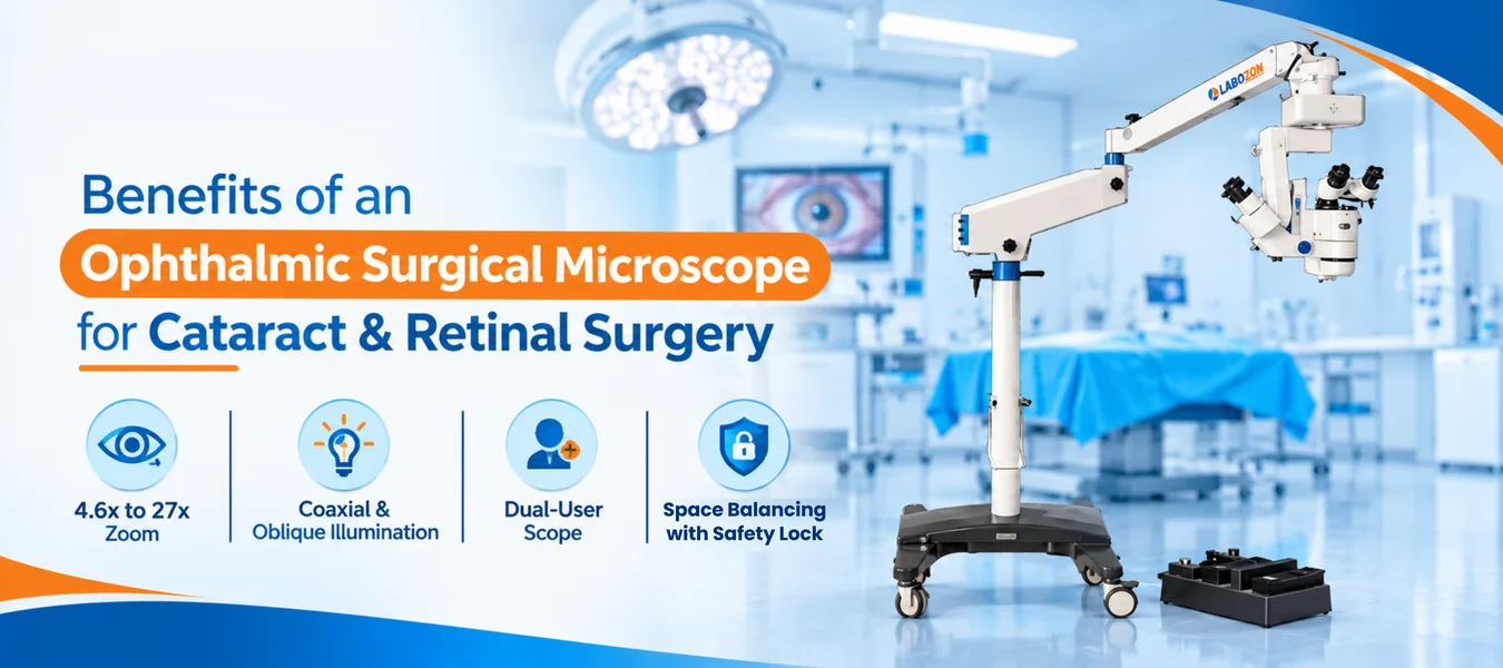
Eye surgery works on structures measured in fractions of a millimeter, and the surgeon's view is the whole workspace. An Ophthalmic Surgical Microscope supplies that view with controlled magnification, adjustable light and a stand that holds its position. This guide explains how the instrument works, how teams use it in cataract and retinal cases and what to review in the Labozon Ophthalmic Surgical Microscope LZ-OPM-A100.
How Optics and Light Work Together
An ophthalmic microscope combines an objective, a zoom system and binocular eyepieces with its own light source. The zoom changes magnification without changing the working distance, so the surgeon keeps the same space above the eye.
In operating microscope ophthalmology setups, light travels along two paths. Coaxial light runs through the same axis as the optics and produces the red reflex, while oblique light enters at an angle and adds depth to the field.
Where the View Matters Most
Ophthalmic surgical microscope use centers on two large areas of eye care. In cataract surgery, teams rely on a stable red reflex to see the capsule edge and lens material during incision, capsulorhexis, lens removal and implant placement.
Retinal work asks for more depth and steadier focus, because vitreoretinal teams handle thin membranes and small vessels. Many ophthalmic surgical techniques share the same needs: a bright field, controlled magnification and a stand that does not drift.
What Each Control Adds in the Operating Room
Continuous Zoom
Surgeons move from a wide field to fine detail without swapping eyepieces.
Adjustable Light
Coaxial and oblique beams are set independently across nine levels.
Dual-User View
A secondary microscope lets an assistant or trainee follow each step.
Steady Positioning
A balanced arm and safety lock hold the head where the team sets it.
Key Details of the LZ-OPM-A100
The Labozon Ophthalmic Surgical Microscope LZ-OPM-A100 is a floor-standing unit on a wheeled base. Its published details are below.
| Feature | Specification |
|---|---|
| Main microscope zoom | 4.6x to 27x continuous, motorized and manual |
| Eyepiece | 12.5X |
| Secondary microscope | 6X, 10X, 16X |
| Field diameter | 46 mm to 8.5 mm |
| Working range | 170 mm |
| Diopter adjustment | ±5D |
| Illumination | 12V/100W halogen cold light, coaxial and oblique |
| Light control | Nine levels, panel control |
| Fine focus and X/Y | 2 mm/s or slower, 50 mm travel |
| Power | AC 220V/110V ±10%, 50/60 Hz, 170 VA |
| Gross weight | 215 kg |
Surgical Model and Slit Lamp Compared
The table below sets two common ophthalmology microscope types side by side using typical features.
| Factor | Slit Lamp | Surgical Microscope |
|---|---|---|
| Setting | Clinic | Operating room |
| Patient position | Seated | Lying down |
| Magnification | Stepped | Continuous zoom |
| Light path | Slit beam | Coaxial and oblique |
| Typical fit | Examination | Surgery and training |
Selection Mistakes That Cause Rework
- Comparing an ophthalmic operating microscope on magnification range alone and ignoring light control.
- Skipping a check of room height and floor space for a stand with a long arm.
- Overlooking assistant viewing needs for teaching cases.
- Leaving the electrical supply unchecked against the voltage the unit expects.
- Asking each supplier for different figures, which makes offers hard to compare.
Teams that want to compare similar stands can start with the operating microscope category before shortlisting.
User Tips and Recommended Practices
- Balance the arm first, then engage the safety lock.
- Set diopters and pupillary distance for each user before the case.
- Start with moderate light and raise it only as far as the case needs.
- Use coaxial light for the red reflex and oblique light for depth.
- Test focus and X/Y movement before draping.
- Place the sterile drape without tugging the optics or the arm.
- Clean lenses gently with lens tissue and approved cleaner.
- Keep a spare halogen lamp near the unit.
- Log lamp hours, cleaning dates and checks.
Exploring Wider Operating Microscope Options
The LZ-OPM-A100 belongs to the operating microscope category, which covers ophthalmic, neurosurgical, dental and ENT stands in several configurations. Buyers usually compare zoom range, illumination type, assistant viewing, arm reach, height range and fine motion control.
The full Labozon operating microscope range shows these stands together, and the Labozon home page opens the other laboratory and medical equipment groups.
See the full specification of the Ophthalmic Surgical Microscope LZ-OPM-A100.
Explore Ophthalmic Surgical MicroscopeRelated Products

Looking for ophthalmic surgical microscope?
Compare available models, review product specifications, and contact the Labozon team for assistance in identifying equipment that matches your laboratory requirements.