
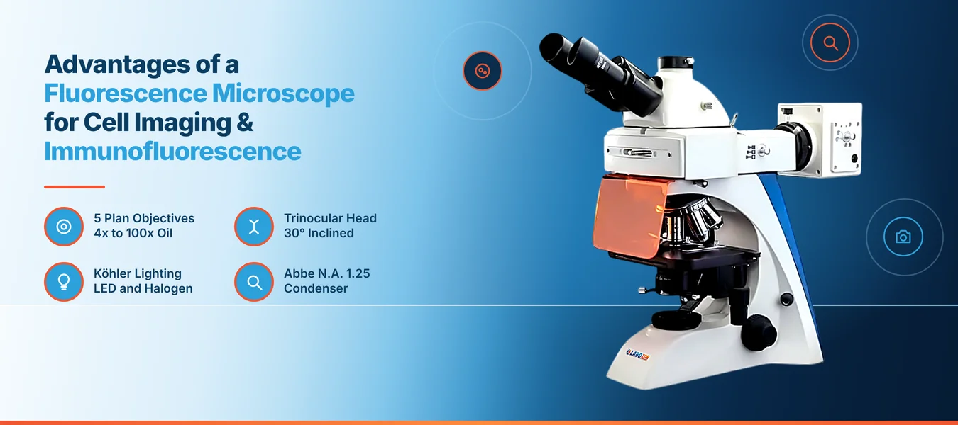
A stained cell under bright light shows shape, but it hides where a specific protein sits. A fluorescence microscope makes labeled targets glow against a dark background, so research, clinical and hospital laboratories can see structures that ordinary optics miss. This guide explains how it works, where it is used, and what to review in the Labozon Fluorescence Microscope LZ-FM-A100.
Fluorescence Microscope Principle
The fluorescence microscope principle is simple. Light of a chosen wavelength excites a fluorescent dye attached to a target. The dye emits light at a longer wavelength, and filters separate that emitted light from the excitation light.
In an epi-fluorescence design, the same objective delivers the excitation light and collects the emitted light. A fluorescence microscope easy diagram follows this path: light goes down through the objective, the dye glows, and the signal travels back up to the eyepiece or camera.
Why Labeled Targets Change Cell Imaging
Fluorescence microscope advantages appear quickly in daily work, because each label marks a specific structure, so nuclei, membranes and proteins can be told apart in one sample.
Specific Labels
Each dye marks a defined target.
High Contrast
Targets glow against a dark field.
Multiple Targets
Different dyes mark different structures.
Image Capture
A camera attachment records still images and live video.
Immunofluorescence at the Bench
Immunofluorescence uses antibodies carrying fluorescent labels to show where a protein sits inside a cell or tissue. A fluorescence imaging microscope makes the stained result visible, and a camera records it for review.
Typical steps include fixing cells, applying primary and labeled secondary antibodies, mounting the slide and imaging with matched filters. The match between dye and filter set matters most, because a fluorescence microscope can only show a signal when its excitation light and emission filters suit the dye in use.
Fluorescence Microscopy Applications
Fluorescence microscopy applications cover many fields. The LZ-FM-A100 page lists advanced research in pathology, cytology, genetics, environmental science and biotechnology.
- Cell imaging and counting
- Immunofluorescence staining checks
- Tissue and pathology review
- Genetic and cytogenetic studies
- Environmental sample screening
These fluorescence microscope uses sit alongside a biological microscope used for routine bright-field checks, and a laboratory microscope of this type often shares bench space with it.
Key Details of the LZ-FM-A100
The Fluorescence Microscope LZ-FM-A100 is an epi-fluorescence unit with a trinocular head and lists these published details.
| Feature | Specification |
|---|---|
| Viewing head | Siedentopf trinocular head, inclined at 30°, 360° rotation |
| Interpupillary distance | 50 to 75 mm |
| Eyepiece | WF10X/20 mm |
| Objectives | Infinite plan, 4x, 10x, 20x, 40x and 100x oil |
| Stage | Double-layer mechanical stage, travel 76 × 52 mm |
| Condenser | Abbe N.A. 1.25 with iris diaphragm |
| Illumination | Built-in Köhler system with 3W LED and 6V/20W halogen, brightness adjustment, field diaphragm |
| Focusing | Coaxial coarse and fine knobs |
| Optional additions | Dark field equipment, phase contrast equipment, C-mount camera adapters, software with camera driver, Original OMEGA filters |
The page does not state which filter sets, dye channels or fluorescence light source come with the unit, so confirm these with Labozon for your dyes.
Fluorescence and Bright-Field Viewing Side by Side
The table shows how the two common viewing modes differ.
| Factor | Fluorescence Imaging Microscope | Bright-Field Microscope |
|---|---|---|
| What is seen | Labeled targets glowing on a dark field | Stained or natural contrast |
| Suited to | Specific proteins and structures | General morphology |
| Sample preparation | Fluorescent labels required | Often simpler staining |
| Care needed | Limit exposure to reduce signal fading | Routine cleaning |
Selection Mistakes That Cause Rework
- Buying on magnification alone and ignoring filter availability for the dyes in use.
- Overlooking the camera port and adapter options when image capture is required.
- Skipping a check of illumination control and field diaphragm adjustment.
- Comparing fluorescence microscope manufacturers on appearance without reviewing optics and accessory range.
- Forgetting stage travel and condenser details for the slide formats used.
Teams reviewing options can browse the microscope category to match optics, head type and camera options to the imaging task.
User Tips and Recommended Practices
- Match the filter set to the dye before each session.
- Limit exposure time to reduce fading of the fluorescent signal.
- Include a control slide without the primary antibody to check background.
- Image in a dim room so faint signals are easier to see.
- Wipe the oil objective gently after each use with lens tissue.
- Store stained slides in the dark and image them soon after preparation.
- Start with a low-power objective to find the field, then move up.
- Record dye, filter, objective and exposure for each image.
Exploring the Wider Microscope Range
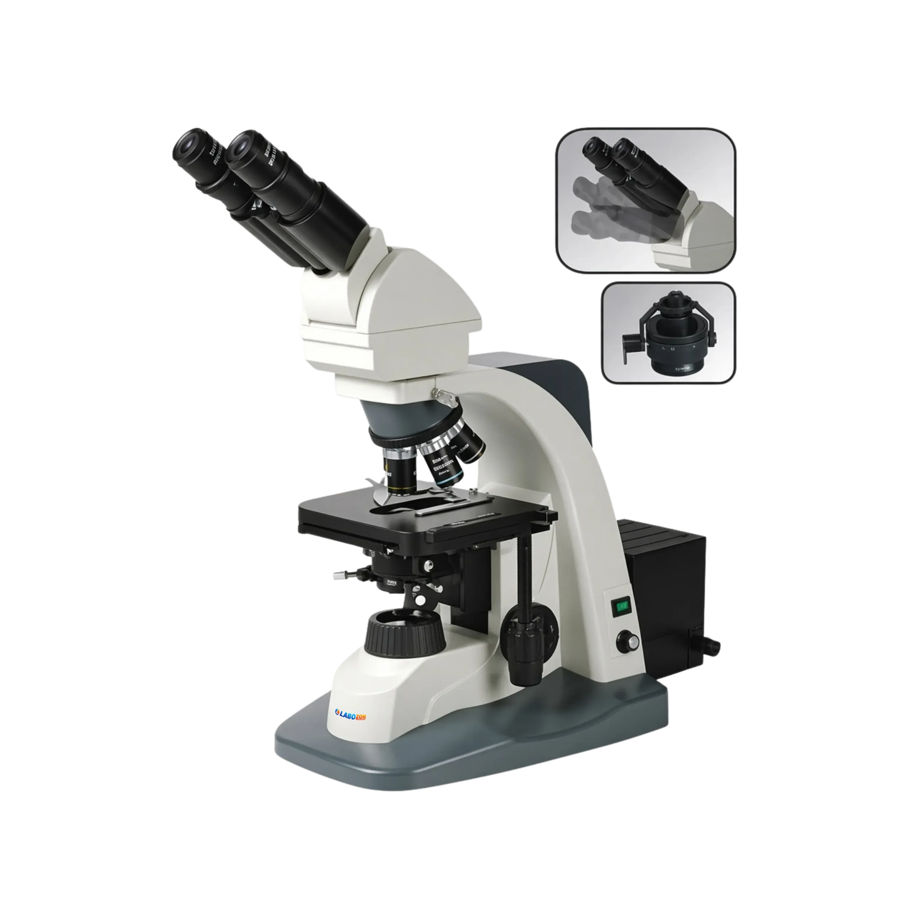
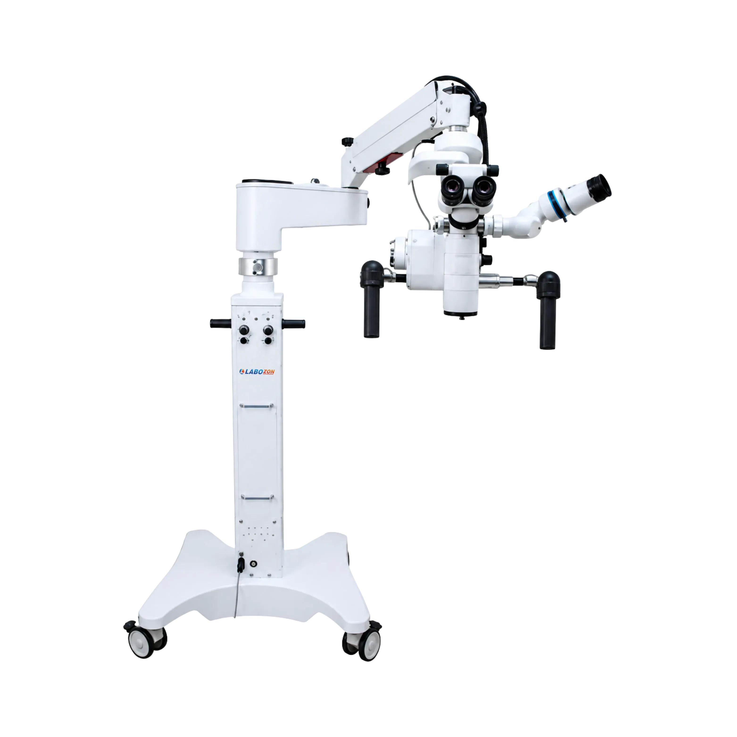
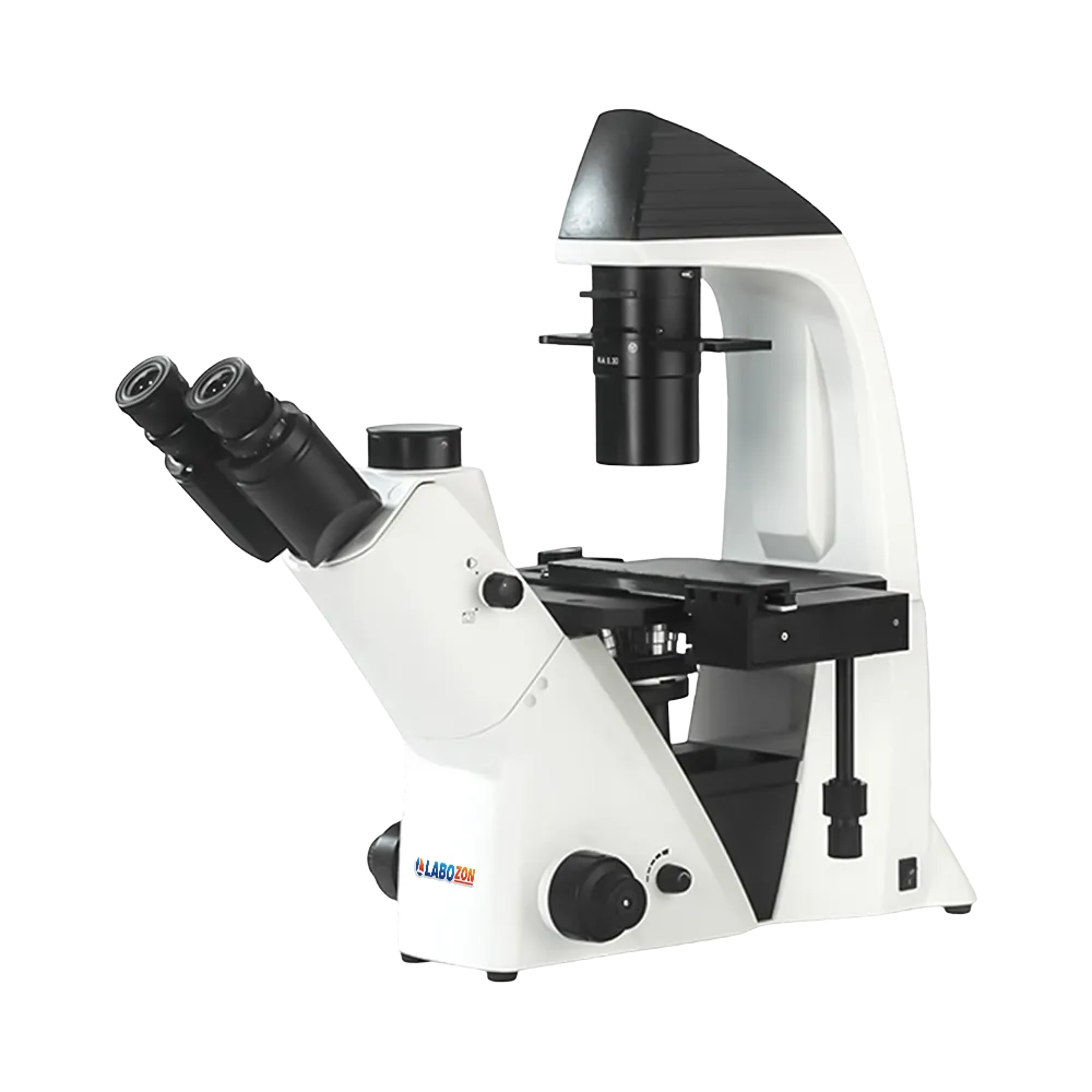
The LZ-FM-A100 belongs to the microscope group, which covers biological, stereo, inverted and fluorescence instruments for research and clinical use. Buyers usually compare head type, objective range, illumination, condenser, stage design and camera options.
The full range of microscopes shows these instruments together, and the Labozon home page opens the other laboratory equipment groups.
See the full specification of the Fluorescence Microscope LZ-FM-A100.
Explore Fluorescence MicroscopeRelated Products


Looking for fluorescence microscope?
Compare available models, review product specifications, and contact the Labozon team for assistance in identifying equipment that matches your laboratory requirements.