
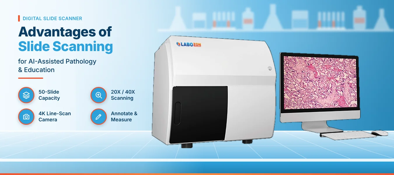
A glass slide can be viewed by one person at a time, and it fades, breaks and travels slowly. A digital slide scanner turns each slide into a detailed image that a team can open, annotate, measure and share. This guide explains how scanning works, why it matters for analysis and teaching, and what to check when evaluating the Digital Slide Scanner LZ-DSS-A101.
From Glass Slide to Whole Slide Image
A scanner captures an entire tissue section, not a single microscope field, and stitches the capture into one large file often called a whole slide image. Reviewers zoom, pan and compare regions on screen without touching glass.
Catalogs use several names for the same idea. A pathology slide scanner and a digital pathology slide scanner both describe an instrument that feeds the wider imaging workflow, so within any digital pathology system the quality of each scan shapes each later step.
How Line Scanning and Focus Work
The LZ-DSS-A101 uses line scanning. A 4K line scan camera with a frequency of up to 66 kHz sweeps across the specimen in strips, and software joins the strips into one image.
Automatic and manual focus modes are listed, and a Z-stack range of 0 to 1.6 mm lets the scanner capture layered tissue. Depth of field fusion then merges the layers into one sharp image.
Where Scanning Helps AI-Assisted Analysis
Image analysis software that counts cells, measures stain intensity or flags regions of interest performs well when scans share the same color and sharpness. High-fidelity color restoration helps keep stained slides consistent from batch to batch.
The product page lists immunohistochemistry applications such as PD-L1 and Ki67 analysis and quantitative research analysis. It does not name specific AI software, so a laboratory should confirm which analysis tools it plans to connect.
Advantages in Teaching and Education
Teaching sets made from glass wear out and drift in color. Scanning a slide once lets an instructor share the same image with a whole class.
Shared Sets
One image opened by a whole group.
Eyepiece View
Microscope simulation with switchable magnification.
Annotation
Instructors mark features directly on the image.
Remote Teaching
Groups learn without sharing one room.
Inside a Digital Pathology Laboratory
A digital pathology laboratory uses scanning for routine work as well as teaching. The listed applications include pathological section scanning, digital slice review and remote diagnosis support in diagnostic labs.
Cytology labs scan TCT slides, immunohistochemistry labs scan IHC slides, and tissue labs scan puncture sections, TMA chips and large tissue sections. Research labs use the images for measurement and key area scaling.
Specifications of the LZ-DSS-A101
The Labozon Digital Slide Scanner LZ-DSS-A101 is a batch-loading scanner for bright field slides. Its published details are below.
| Feature | Specification |
|---|---|
| Capacity | 50 slides, automatic loading |
| Magnification | 20X and 40X |
| Objective | 20X Plan Apochromatic lens, NA 0.8 |
| Pixel size | 0.38 µm at 20X; 0.19 µm or better at 40X |
| Scan time | 25 seconds or less for a 15 mm x 15 mm area at 20X |
| Camera | 4K line scan camera, up to 66 kHz |
| Focus | Automatic and manual; Z-stack 0 to 1.6 mm |
| Label reading | Barcode reader |
| Sample types | H&E, IHC, frozen section, special stain and immunocytochemical slides |
| Workstation | Intel i7 or higher, 3 TB hard disk, 23.8-inch 2K display, Windows 10 |
Glass Review Compared With Scanned Review
The table below sets the two review routes side by side.
| Factor | Glass Slide | Scanned Image |
|---|---|---|
| Readers at once | One | Many |
| Annotation | Marker on glass | Digital, reversible |
| Measurement | Manual | On-screen tools |
| Color over time | Fades | Fixed in the file |
| Sharing | Courier | File transfer |
Selection Mistakes That Cause Rework
- Judging by scan time alone and ignoring pixel size.
- Forgetting that scans create large files and storage grows quickly.
- Overlooking the slide types the laboratory actually runs.
- Skipping a test scan on pale or thick sections.
- Comparing digital slide scanner manufacturers without asking about software and service access.
Teams that want to compare imaging instruments can start with the microscope category before shortlisting.
User Tips and Recommended Practices
- Clean slides and remove dust and fingerprints before loading.
- Check coverslips for bubbles, since bubbles blur the scan.
- Label each slide clearly so the barcode reader can identify it.
- Run a test scan on pale and thick sections before a full batch.
- Select the scan area carefully to avoid empty glass and large files.
- Use the same settings for slides stained in the same batch.
- Back up scanned images on a schedule and plan storage growth.
- Review a few scans at full zoom before sending a batch for analysis.
- Keep notes on settings so results stay comparable.
Exploring Wider Microscopy and Imaging Equipment
The LZ-DSS-A101 sits in the microscope category, which groups microscopes and related imaging instruments for sample viewing. Buyers usually compare magnification range, image quality, throughput, software tools and workstation needs.
The full Labozon microscope range shows these instruments together, and the Labozon home page opens the other laboratory equipment groups.
See the full specification of the Digital Slide Scanner LZ-DSS-A101.
Explore Digital Slide ScannerRelated Products


Looking for digital slide scanner?
Compare available models, review product specifications, and contact the Labozon team for assistance in identifying equipment that matches your laboratory requirements.